An ASU professor and her team are currently discovering new and more accurate ways of measuring electrical activity in the human brain.
This research could increase understanding of the electrical activities in the human body and brain, as well as helping to quickly diagnose neurological problems patients may have in non-invasive and accurate ways.
School of Biological and Health Systems Engineering associate professor Rosalind Sadleir and assistant research professor Munish Chauhan, along with biomedical engineering graduate research associate Sulagna Sahu, are part of the research team led by Sadleir.
The team's research has been funded through multiple grants, three of which came from the National Institutes of Health.
One of their studies is using an MRI to visualize and record electrical currents from neuromodulation. The other study they are working on involves developing a new functional imaging technique in which brain activity can be measured through different stimulations.
How the research works
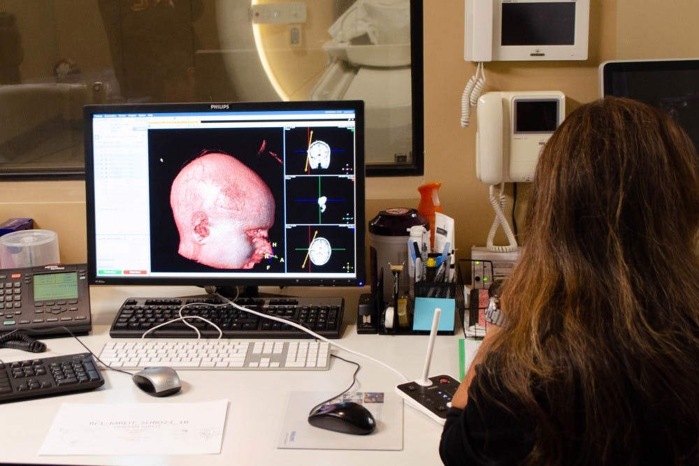
According to Sadleir, health professionals in the past — and even now — have used invasive measures, like removing pieces of the brain, to measure the brain's electrical currents with computers and models. Her team's new research allows them to measure the whole brain and its electrical currents without surgery.
“We have the ability to image where the current is flowing and pick out our own individual subject's properties, the things that determine where the current flows,” Sadleir said. “If we know precisely where the current is flowing, we can begin to think about getting some information about what is the connection between the stimulation and the effect.”
This is the first time the team is using this kind of technology on humans and testing the results, Sadleir said.
A new type of brain imaging
The team is also developing a new technique for functional imaging, which gives them the ability to see where electrical activity is occurring in the brain, Sadleir said.
Typically, this is done through a process called blood-oxygen-level-dependent imaging, or BOLD fMRI. When part of the brain is active, energy is used and blood comes into the areas that have been stimulated to replace the energy, which the BOLD fMRI detects.
The new procedure they are working on uses a functional MRI that they hope will be more accurate than the current process.
“We’re actually measuring signals from the brain cells themselves,” Sadleir said. “Blood comes along and there is a time delay, so there's a few second delay between activity starting and blood coming in — we hope our method will be more direct and more precise.”
In this particular study the team uses a 3-back memory test where they give subjects a list of letters, and they press a button when they see the letter repeat. Each test has a different stimulation, ranging from none at all to a certain level of electrical stimulation. The stimulation is given through electrodes which are meant to affect a subject's memory.
Taking images of the skull in an MRI
Sadleir recently completed a separate task that involves imaging the skull in an MRI, and she has applied for a separate grant to fund that research as well.
Before, a skull in an MRI scan would typically just look black, but the new MRI ultrashort echo time imaging can make a patient-specific map of the skull.
Next, Sadleir and her team will compare the new method to the old and examine differences in the results.
“It's generally been difficult — if not impossible — to measure the skull,” Sadleir said. “Any signals developed in the skull have gone away by the time we measure it, it's much too quick.”
All the work done in the laboratory is done at the Barrow Neurological Institute, combining both Barrow's and ASU’s experience and expertise. The work can help MRI usage become more effective and make it easier to find health problems.
“The amazing thing about MRIs — compared to other imaging systems — is it gives you such subtle images that you can pick out some really interesting processes happening non-invasively,” Sadleir said. “Subtle changes in magnetic properties that are related to changes in a disease or in our case, electrical activities, it keeps on evolving all the time.”
What comes next
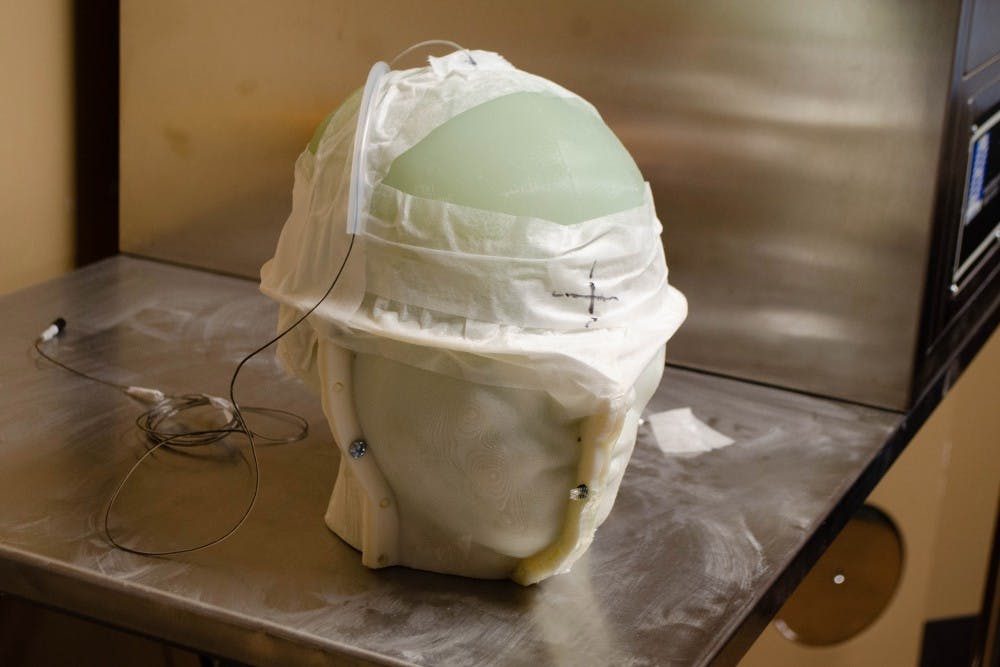
The team's work does not stop there. They are also conducting research on an even more accurate — but invasive — way to detect electrical activities using a model of the brain, which they call a phantom.
Chauhan said they are working on testing a deep brain stimulation procedure. Here, an electrode is placed inside the subject's brain and can target a specific area and directly measure the activities that occur there. A connector controls four different parts on the electrode that sends an electrical current when activated.
Take Parkinson's disease for example, Chauhan said, often subjects with the disease experience shaking in their hands. In this procedure, the electrode could be placed into a direct spot in the brain, and an electrical current could be used to stimulate it and stop the hands' shaking.
Though this procedure is highly effective, it is more invasive than others. It requires surgery and an electrode being placed directly in the brain that can not be done in an MRI.
“Right now we are trying to get the electrical activity of the living tissues without any surgery,” Chauhan said.
Applying the data collected by the MRI
Sahu said she is working on taking this MRI data, putting it into a software and simulating different conditions to the neuron.
Right now she is working on the trigeminal nerve in the brain, which is responsible for the sensations you get on your face, to hopefully show the electrical activity that is activating it. She can also show the electrical current's starting point is from the team's electrode, which can potentially help with memory tasks.
“I can take one data set and turn it into what I need," Sahu said. "I can exactly simulate the experiment into the computer.”
Sadleir and her team plan on continuing their research with the goal of increasing their understanding of electrical activity in the human brain, which can lead to faster neurological diagnoses.
Correction: A previous version of this story confused Sahu with another member of the research team. A previous version of this story also miscategorized what phase in the grant process Sadleir’s skull imaging process was in and misstated Sadleir’s title. The story has been updated to reflect those changes.
Reach the reporter at wmyskow@asu.edu and follow @wmyskow on Twitter.
Like The State Press on Facebook and follow @statepress on Twitter.

Wyatt Myskow is the project manager at The State Press, where he oversees enterprise stories for the publication. He also works at The Arizona Republic, where he covers the cities of Peoria and Surprise.


