At ASU's Biodesign Institute in building C, there's a 3D-printed model of a machine in the basement. Signs around the model say "fragile" and "do not touch." But Mark Holl picks up a piece of it anyway, because he made it himself.
The device is called the compact X-ray light source. CXLS is a prototype, and experiments are still being run to ensure its stability and to perfect a different model, the the compact X-ray free electron laser.
Holl led the team that designed the CXLS' housing facility, and is now the chief engineer on the CXFEL project at ASU. The goal of the lab is to perform experiments on microscopic, dynamic samples, resulting in moving images on the atomic level.
It hit a major milestone in February when scientists generated the first X-rays. Just a month later, the project was awarded a $90.8 million research award from the National Science Foundation, the largest NSF research award in ASU history.
Before descending the stairs to see the device, Holl explains how it works using the model. He picks up the photoinjector, the yellow piece at the head of the model, and begins to demonstrate.
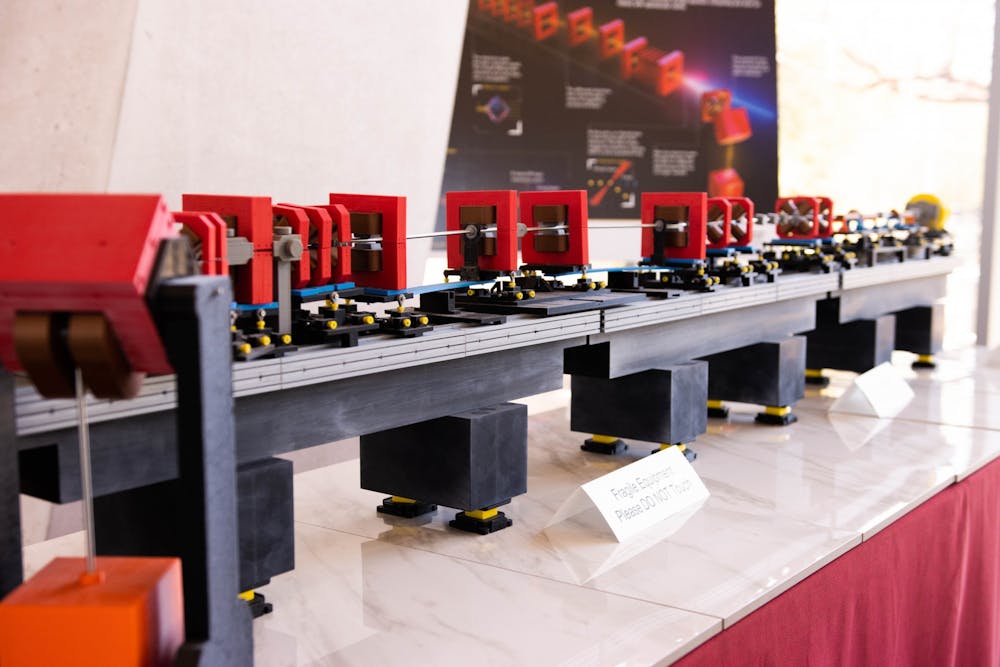
"What happens is the laser, or UV laser, comes in and hits the photocathode, a copper surface," he said. "There's something called a photoelectric effect, Einstein got a Nobel Prize for this, and if you hit with a UV beam — puff — off comes a burst of electrons."
The electrons are then caught in an electric field with nothing but a vacuum to slow them down, and are accelerated close to the speed of light. The electrons travel through a series of magnets that steer and direct the beam to the interaction point.
The energy density of the electron beam has to be lowered before it is bent and thrown away.
"When those electrons are bending around, we cross over at a point of tangency to the arc. Wherever the electrons are going when we cross over the laser beam, off come the X-rays, and they go at the tangent to the electron beam direction," said Holl. "We pull off those X-ray pulses, and the X-ray pulses are very special, and very pure, and very short. When they're very short, then the X-rays can go into a sample and be gone."
The electrons are then put into a dump, and the X-rays are sent to image the molecules in the experiment being performed. Meanwhile, the radiation produced during the experiment is contained to the vault, thanks to a six-foot concrete foundation and a built-in Faraday cage.
In a March 14 meeting with The State Press, ASU President Michael Crow said the instrument is "further evidence of that (the University's) competitiveness. That grant (from NSF) had to be voted on by the National Science Board."
"We had already invested $80 million in the project including special rooms that had to be built over there," he said. "If you go over there, the walls are like three-foot-thick, and they're filled with lead because you can't let those lasers get out the door to the core of the planet and then, you know, Thor comes out of there."
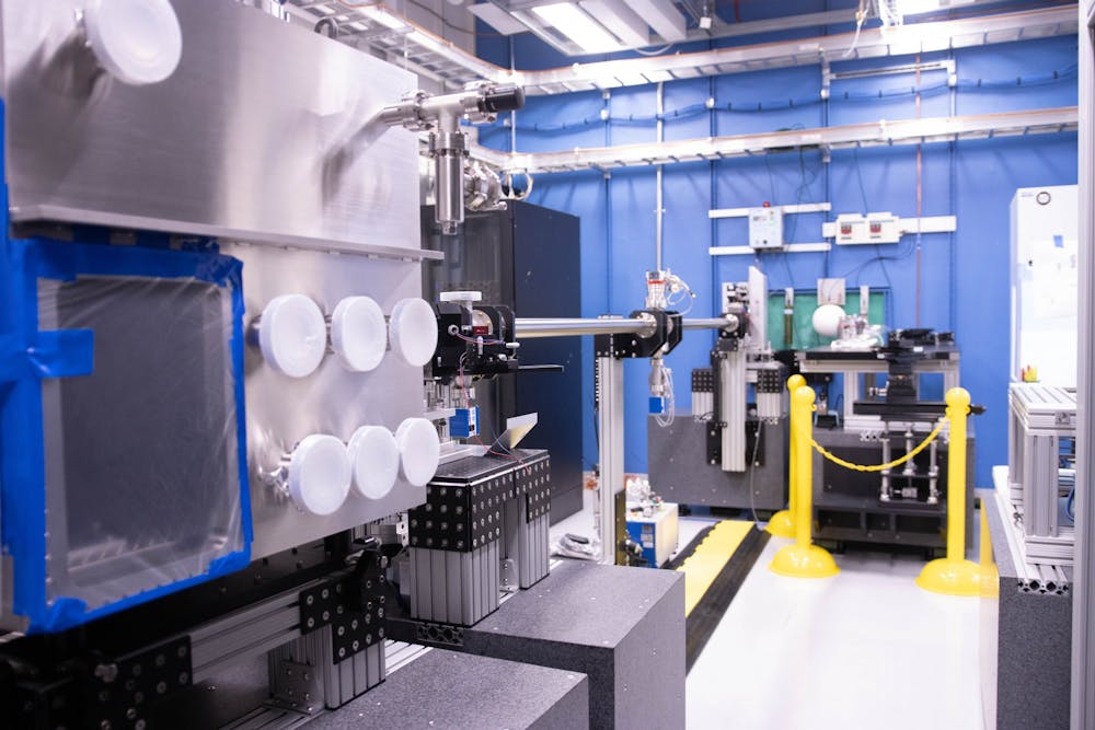
Holl leads us down a long flight of stairs to the floor that houses the CXFEL labs. It's a unique feat that might be overlooked if you are unfamiliar with X-ray light source technology. Similar systems in other locations are built around 30 feet underground and run for miles. CXLS takes up about 5,000 to 6,000 square feet, in comparison.
The CXLS and the later model, the CXFEL, won't replace larger X-ray lasers. Instead, they will expand light source imaging into a new niche, giving researchers clearer dynamic images of microscopic samples than ever before.
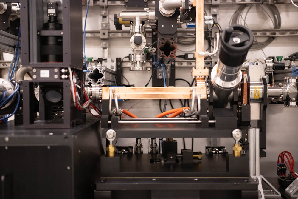
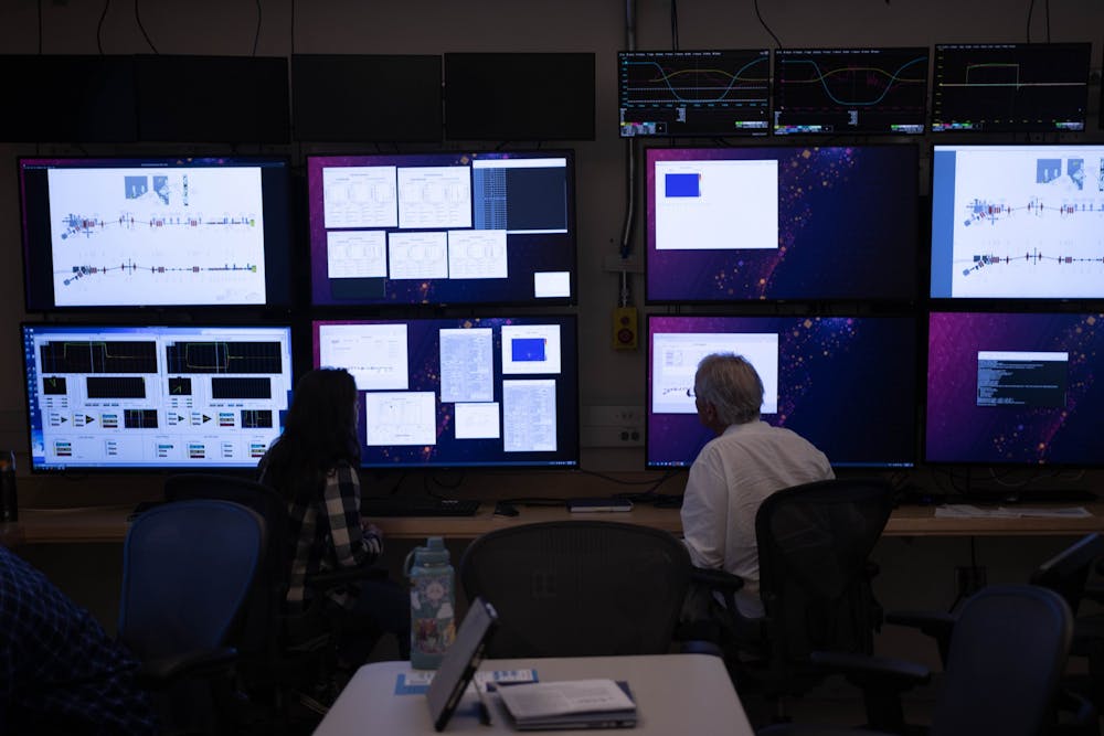
The first room Holl shows us is the Hutch 1 control room, where William Graves, the project's chief scientist, and students are running and testing the stability of the beam. To do this, the room's many monitors are dedicated to live data feeds and control boards.
"Earlier today, we got one of our big parts back, and it's working again. We're at high energy right now, and we are bringing the electron beam back up to full operational," said Taryn Brown, a graduate lab and research assistant studying materials science and engineering.
Brown and Graves are monitoring what's called a scintillator screen, which glows when electrons hit it. They make adjustments to the placement of the beam by changing the machine's various magnets.
On the other side of the control room's wall is the Hutch, where researchers run experiments.
The lab was designed in 2016, and built between 2017 and 2018. Holl said the process was like "building an airplane in mid flight." The six feet of concrete that support most of the lab were poured by a hundred cement trucks in one night, Holl said.
Moving to the vault, where the majority of CXLS is housed, the team in the control room stops running the beam, and after a scan of the room's radiation levels for safety, Graves shows us the instrument up close.
Pointing out the accelerator and magnets he explains, "The copper part (the accelerator) changes the energy. In physics terms, we would say that the copper part does the work because the change in energy and magnetic fields famously can't do any work, so they can't accelerate or decelerate, but they can change direction."
When he was an undergraduate and first introduced to accelerators, Graves thought it was neat that they had progressed beyond problems in the back of a physics textbook. Now, he's looking forward to working on the improved CXFEL model.
"The CXFEL, that has never been built before, and it's pretty exciting," Graves said. "It'll look exactly the same. The same three pieces of copper and yellow magnet and red magnets, but all but the guts of it will be a little different, and there's going to be subtle changes to the shapes inside the copper."
Later this year, the lab intends to begin accepting proposals for research using the CXLS. According to Graves, the machine may be powerful enough to watch reactions on an atomic scale.
"In biochemistry, it's going to take movies of molecular reactions, and then in quantum materials, there are these really subtle effects that happen with the fully coherent light that we're going to make," Graves said. "We think that we can probe them in ways that's never been done, and we'll be at the quantum limits of what's possible."
Edited by Annie Graziano, Reagan Priest and Piper Hansen.
Reach the reporter at aeagerto@asu.edu and follow @audrey_eagerton on Twitter.
Like The State Press on Facebook and follow @statepress on Twitter.
Audrey is a senior studying journalism and mass communication. This is her fifth semester with The State Press. She has also worked at The Arizona Republic.