 Rosalind Sadleir, assistant professor at the School of Biological and Health Systems Engineering, poses for a portrait at the Interdisciplinary Studies building on the Tempe campus on Nov. 17, 2014. Dr. Sadleir has worked with students and faculty on research for over 10 years to develop a new way to see electrical activity as it occurs in the brain. (Photo by Andrew Ybanez)
A new way to see electrical activity as it occurs in the brain will be the product of years of hard work by students and faculty members at ASU.
Rosalind Sadleir, assistant professor at the School of Biological and Health Systems Engineering, has dedicated nearly 10 years of her career as a biomedical engineer and educator to developing a better method of measuring brain activity.
Sadleir said she’s collaborated with other SBHSE faculty members, including Vikram Kodibagkar, and SBHSE students to develop a more precise, quantifiable way to observe the brain’s electrical circuitry. She’s also collaborated with researchers in South Korea.
“We’re hoping that after we’re properly up and running, that we’ll be able to use this sort of technique to investigate anything,” she said. “It will help us in understanding connectivity between different parts of the brain, and it will help us to get a much deeper understanding of how one thing connects to another.”
Sadleir’s research has not yet been used in humans, but she and others who work in her lab have been working extensively with samples of neural tissue taken from Aplysia Californica specimens, a type of sea slug known commonly as the California sea hare.
Sadleir said she expects that her research will eventually be used to study the human brain, and it will enable scientists like her to learn more about Alzheimer’s disease, temporal lobe epilepsy and Parkinson’s disease.
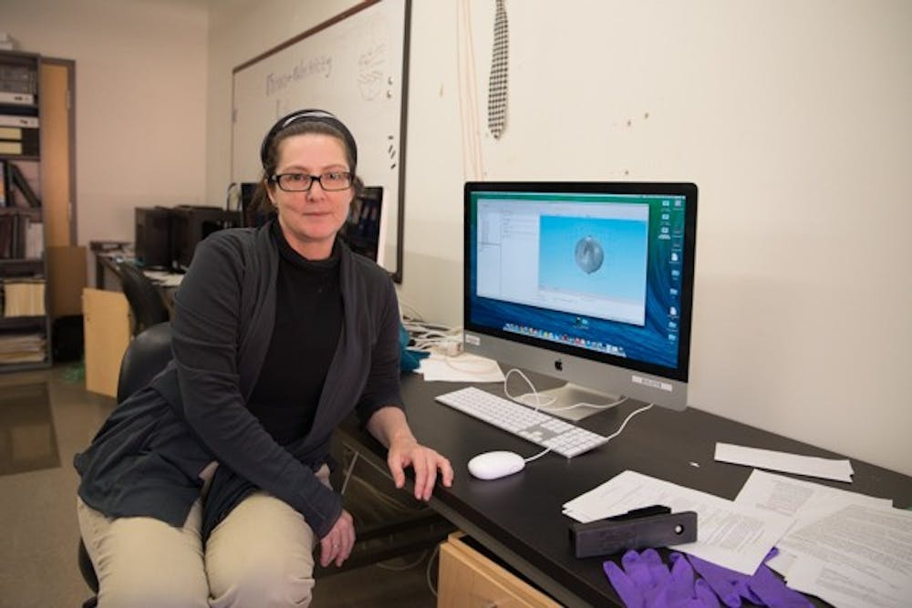
 Rosalind Sadleir, assistant professor at the School of Biological and Health Systems Engineering, displays a computer program used for research at the neuro-electricity lab on Nov. 17, 2014. Dr. Sadleir has worked with students and faculty on research for over 10 years to develop a new way to see electrical activity as it occurs in the brain. (Photo by Andrew Ybanez)
She uses a technique called magnetic resonance electrical impedance tomography, which uses “electrical contrasting” to indicate electrical activity in MRI images of the brain.
Sadleir said this technique is made possible because the electrical properties of cells change when they are active, leaving them more vulnerable to a “trickle current” that shows up in images produced by MRI machines.
“When a cell produces electrical activity, its conductivity changes,” she explained. “It’s easier for electricity from outside to go through a cell when it is active, as opposed to when it is not active. We can use that fact to interrogate cells.”
By exposing brain cells to a trickle current, Sadleir said she is able to more accurately track and measure activity in brain tissue.
Sadleir’s technique works in contrast to traditional MRI methods, which are only able to identify brain activity as it correlates to blood flow in different regions of the brain.
Most modern-day researchers and medical practitioners observe brain activity by analyzing small changes in MRI images created by the oxygenated blood that accumulates around active areas, Sadleir said.
She said this is a useful way to locate active regions of the brain when it preforms specific tasks, because oxygenated blood flow is generally channeled to areas expending the most energy.
However, Sadleir said her MREIT technique takes brain activity imaging to the next level.
“While (this method) is very useful, it’s not actually directly telling us about the activity,” she said. “It’s just showing us a correlation between blood flow and areas of the brain that are active.”
 Lab materials strewn across a table at the neuro-electricity lab at the ASU Tempe campus. (Photo by Andrew Ybanez)
Aprinda Indahlastari is a graduate student studying biomedical engineering at ASU who has been working in Sadleir’s lab since last September. She said she is amazed at the progress the lab has made improving brain activity imaging since she has been working there.
“I’m fascinated by how much we know now that we didn’t know before,” Indahlastari said.
She said she thinks the work environment of the lab has contributed to its success, calling it both “productive” and “enjoyable.”
Steve Keim, biomedical engineering doctoral candidate in the Sadleir lab, said the work he does is “time-consuming, but rewarding,” but like Indahlastari, he said he enjoyed being involved in Sadleir’s MREIT research.
“If I had to describe this research with just three adjectives, I’d say it’s groundbreaking, it’s exhilarating and it’s interesting,” he said.
CORRECTION: Due to a reporting error, a previous version of this article incorrectly stated the common practices for observing brain activity. This version of this article has been updated with the correct information.
Reach the reporter at megannphillips@asu.edu or follow her on Twitter @megannphillips
Like The State Press on Facebook and follow @statepress on Twitter.


